
- Pediatric Surgery
- Pediatric Choledochal Cyst
Pediatric Choledochal Cyst
A pediatric choledochal cyst is a rare condition present at birth that can lead to complications if untreated. These cysts are abnormal, fluid-filled sacs that develop in the bile ducts that carry bile from the liver to the small intestine. Such cysts can stop or reduce bile flow, leading to liver problems, pancreatitis, and a higher risk of bile duct cancer. They are typically treated with surgery.
What causes a choledochal cyst?
What causes choledochal cysts is unknown. What we do know is they tend to be more common in females and children of Asian descent. It is thought they occur during development in the mother’s womb because of a problem with the formation of the bile ducts. Most cases are diagnosed before age 10, but symptoms can appear years later and in adulthood.
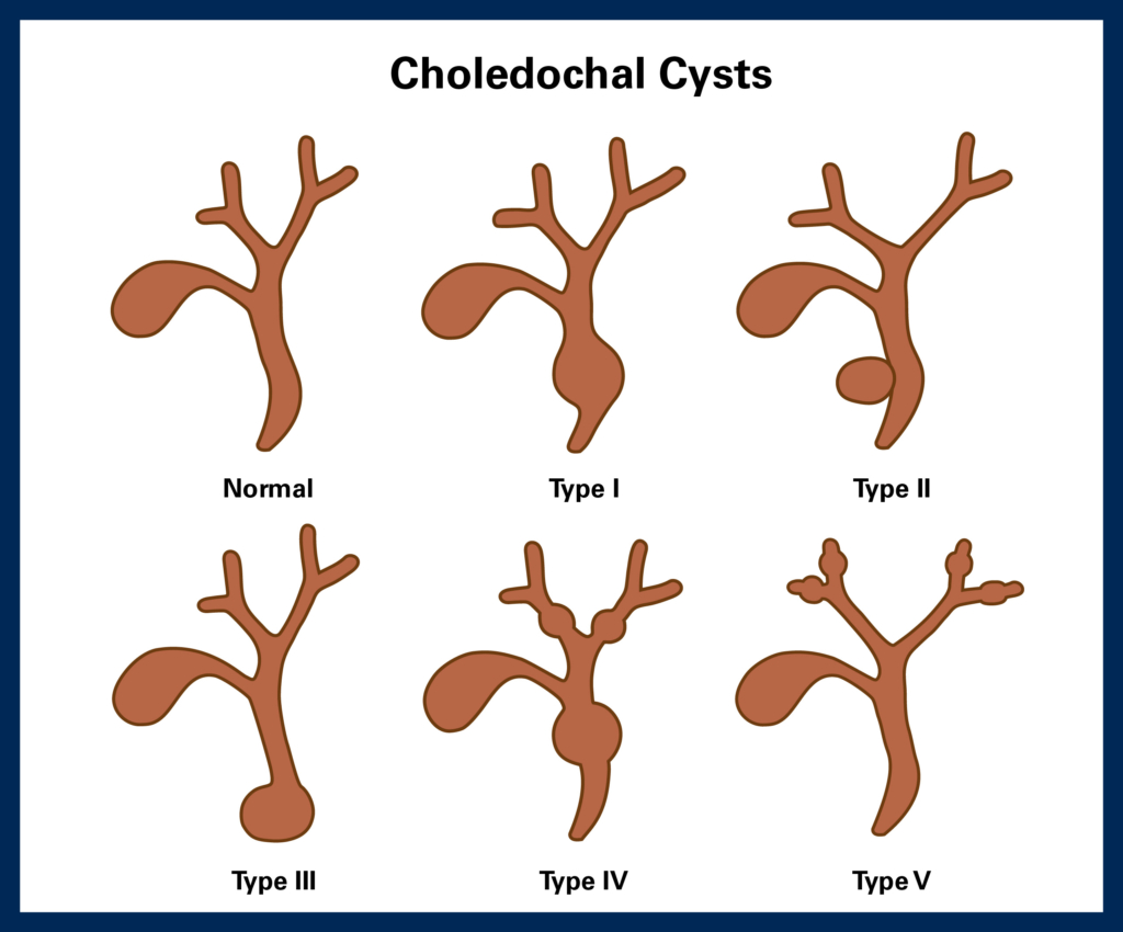
There are several types of choledochal cysts that are based on the shape of the cyst and where in the bile duct it’s located:
Type I: the most common form, characterized by bile duct enlargement outside the liver
Type II: a cyst on the bile duct outside the liver
Type III: a cyst in the pancreas or wall of the duodenum (small intestine)
Type IV: a cyst in the liver along the bile ducts
Type V: several cysts of the bile duct inside the liver, also known as Caroli disease
What are the symptoms?
Common symptoms of choledochal cysts include:
- Abdominal pain that may come and go
- Jaundice, which is characterized by yellowing of the whites of the eyes and skin
- Nausea
- Vomiting
- Fever
- A mass in the abdomen that can be felt through the skin
- Pale stool or dark urine
How is it diagnosed?
Choledochal cysts are sometimes detected before birth during a pregnancy ultrasound. In other cases, a diagnosis is made following a physical exam, review of medical history, and blood tests. If a choledochal cyst is suspected, imaging studies may be ordered, such as a CT scan, magnetic resonance imaging (MRI), cholangiography with X-ray and ultrasound, hepatobiliary iminodiacetic acid scan (HIDA), or endoscopic imaging.
How choledochal cysts are treated
Choledochal cysts are treated surgically, performed with a traditional open procedure involving a large incision in the skin to access organs and tissues, or laparoscopically, a minimally invasive surgery where small incisions are made and a camera inserted to guide the surgeon. Both types of surgery are done with the patient under general anesthesia. The cyst or cysts are surgically removed and, if needed, bile ducts reconstructed or repaired with a portion of the intestine.
Most children will stay in the hospital for a few days and can resume normal activities in a few weeks. Yearly follow-ups are recommended to monitor for possible long-term complications in the reconstructed bile duct and the risk of cancer in the other bile ducts.