Imagine being short-winded and experiencing chest pains from just taking the trash to the end of the driveway. It was a wake-up call for Lee Pratt, who was born with a congenital heart disease. Over time, his condition had steadily worsened without resolution. After connecting with a team of cardiac specialists at UT Physicians, Lee received the heart surgery he knew he’d someday need.
Understanding Lee’s heart
Born with multiple heart defects, Lee had a vascular ring repair at 18 months old to correct a flaw that can put pressure on the trachea or esophagus. At the time, it was decided not to treat a small hole in his heart, a ventricular septal defect (VSD), and to monitor it.
Lee didn’t let his condition define his life, however. He played football, baseball, and basketball and ran track in his youth despite regular hospital visits and recurring chest pains.
“I was raised to not accept limitations,” Lee said. “My parents said they could let me live in this bubble or a normal life. I’d rather just accept what I have and go on living normally.”
Lee’s complex congenital heart disease repeatedly landed him in the emergency room with chronic chest pain. He was miserable. He even had to quit his job. While previously considered minor, his symptoms became more problematic and severely impacted his daily activities.
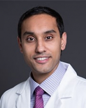
“Lee was a diagnostic dilemma,” said Sukhdeep S. Basra, MD, an interventional and advanced heart failure cardiologist. “Although he was referred to me for VSD closure, his symptoms were out of proportion to the VSD.”
Lee was referred to Basra in late 2023 for a possible VSD repair, but Basra’s analysis suggested there could be other problems with Lee’s heart.
Partnering with a congenital heart disease specialist
Basra collaborated with Santosh C. Uppu, MD, an adult congenital heart disease specialist at UT Physicians Adult Congenital Heart Disease Clinic – Texas Medical Center, for consultation and evaluation. Uppu also believed Lee’s VSD was small and suspected it wasn’t the primary cause of his symptoms. With a CT scan, they evaluated Lee’s VSD, heart function, and coronary arteries. That’s when an anomaly in his coronary artery called a myocardial bridge was detected. It’s a condition where heart muscle fibers compress an artery with each heartbeat, limiting how much blood goes to the heart muscle.
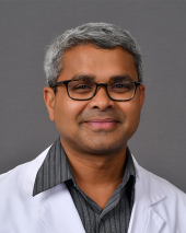
“Lee’s was unique because it was deep – like 4 or 5 millimeters deep, which is significant,” said Uppu, associate professor of cardiology at McGovern Medical School at UTHealth Houston. “The artery was getting squished and compressed over time, causing his symptoms.”
The team presented Lee’s case at one of their cardiac meetings with interventional cardiologists and surgeons. They wanted more investigation.
Investigating with additional testing
Basra and Kiran K. Mallula, MD, a UT Physicians pediatric cardiologist and an associate professor of pediatric cardiology at McGovern Medical School, performed a cardiac catheterization to further measure Lee’s VSD and test blood flow, which confirmed that the VSD wasn’t large enough to explain his symptoms. Basra then performed a special series of invasive tests in the catheterization lab and administered medications to make Lee’s heart work vigorously. He also used a special catheter that uses light to create a 3D picture of Lee’s heart artery and visualize the myocardial bridge from inside the vessel. This confirmed that Lee’s symptoms were coming from the coronary artery compression.
“This is the one of the few times in my career I’ve seen testing confirm such a significant myocardial bridge,” said Basra, associate professor at the Center for Advanced Cardiopulmonary Therapies and Transplantation at McGovern Medical School. “The images were so profound we’ve submitted this as a case report to share at an international cardiology meeting.”
What started as a consultation for a single heart condition turned into a discovery of multiple issues requiring complex surgery.
The power of team-based care
Basra and Uppu immediately contacted Danny Ramzy, MD, PhD, a cardiothoracic surgeon at UT Physicians Cardiothoracic & Vascular Surgery – Texas Medical Center, for a minimally invasive robotic procedure to address Lee’s myocardial bridge, which is Ramzy’s specialty. However, Ramzy’s presurgical evaluation revealed there was more to the story — a problem with Lee’s aortic valve area due to a membrane that turned out to be significant.
“This case emphasizes the importance of looking beyond the initial diagnosis when symptoms don’t quite align with what we expect to see,” Basra said. “Through collaboration and specialized testing, we were able to find the true cause of his symptoms and help him get the treatment he needed.”
The need for open-heart surgery
Instead of a robotic-assisted operation, Ramzy successfully performed open-heart surgery to address three separate issues:
- Closure of the hole in Lee’s heart
- Unroofing of the myocardial bridge (cutting through a muscle band over a coronary artery to improve blood flow to the heart)
- Removal of a subaortic membrane (tissue growth below the aortic value)
“I’m glad I did his surgery in the traditional, open way,” said Ramzy, professor and UTHealth Houston Chair in Cardiac Surgery at McGovern Medical School. “He had a subaortic membrane, a rigid tissue right below the aortic valve, that obstructed blood flow. That’s what I was suspicious about when I looked at the echocardiogram.”
Working toward a quick recovery
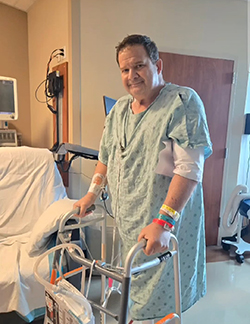
Despite the complexity of the surgery, Lee’s recovery was remarkably quick. It especially surprised his medical team, as he was sitting up in a chair the morning after open-heart surgery.
“I told Dr. Ramzy right before they gave me the anesthesia, ‘You do your job, you tell your people to do their job, and I’ll do mine in recovery,’” Lee said. “I couldn’t let them down.”
Basra followed up on Lee in the ICU after his surgery. Uppu said that a few days after surgery, Lee was all smiles.
“I have never seen anyone recover from heart surgery smiling,” Uppu said. “His burdens and problems were lifted.”
Uppu said Lee thanked the UT Physicians team for identifying and addressing his heart problems.
“Nobody wants to go for heart surgeries because it’s a big procedure,” said Uppu, who works exclusively with congenital heart patients who have had heart surgeries or need them. “Mr. Pratt was asking for it, even though he didn’t know what was causing his symptoms. When we took care of it, he felt like a new person.”
Lee was discharged four days after surgery and was soon participating in cardiac rehabilitation three times a week. Ramzy said he now has a normal life expectancy.
Ramzy credits part of the success to having a good patient partner like Lee who asked good questions and was committed to recovery and rehabilitation.
“He’s had VSD his whole life, so he asked why it needed to be repaired now,” Ramzy said. “He understood what was going on and trusted his physician and the system.”
Looking forward
Basra and Uppu will continue to monitor Lee in follow-up in their specialized clinics. Lifelong follow-up care is recommended for any patient who has a procedure as a child with a congenital cardiac diagnosis. Conditions can change as people age, or issues can develop later in life.
Lee advises others facing similar challenges to be their own advocate if they’re not getting the answers they need. “Doctors have machines to help diagnose you, but if you’re still having problems, let them know,” he said.
Living his new life
Today, Lee is thriving in cardiac rehabilitation, exercising 55 minutes daily, three times a week as part of a 12-week program. He’s lost more than 20 pounds and, most importantly, hasn’t experienced cardiac-related chest pain since his surgery in October 2024.
“My wife says she got her active husband back,” Lee said. “I’ve changed my life completely.”